
District Health Boards, once known as Hospitals.
DHB CEO, earlier named Hospital Superintendent (often a reluctant Doctor Clinician dragged away from patients to run a hospital).
Chief Nursing Officer, formerly termed Matron.
Lead Sector Physiotherapist, previously known as Charge Physiotherapist.
Client, in the past called a Patient.
Purchase Order, formerly named Treatment.
These are a few examples of how medicine has been massaged or even manipulated into a new medical image based on becoming a business model rather than the medical likeness of the past. This image change has not been led by clinicians, but by society, health managers, and politicians, with some success bending the medical ethos into a replica of their own style of service. The current situation with the health system can be assessed by readers as to the efficacy of the new look.
A few years ago, as the new era for a large DHB was signalled to surgeons, the surgeons held an informal gathering to discuss whether to support the new CEO or not. The consensus was they would give the CEO and the proposed systems a chance.
Medical protocols faded, replaced by business procedures. Then, a Memo was distributed to all clinicians: “Biopsies cost X amount of dollars, please only take biopsies that are positive.” Memo to Judges: “ Court Hearings cost X amount of dollars, please only assign cases where the Defendant is to be found guilty. “
The CEO was promoted downwards, probably still being paid well for fine work, but away from all dangers incoming or outgoing.
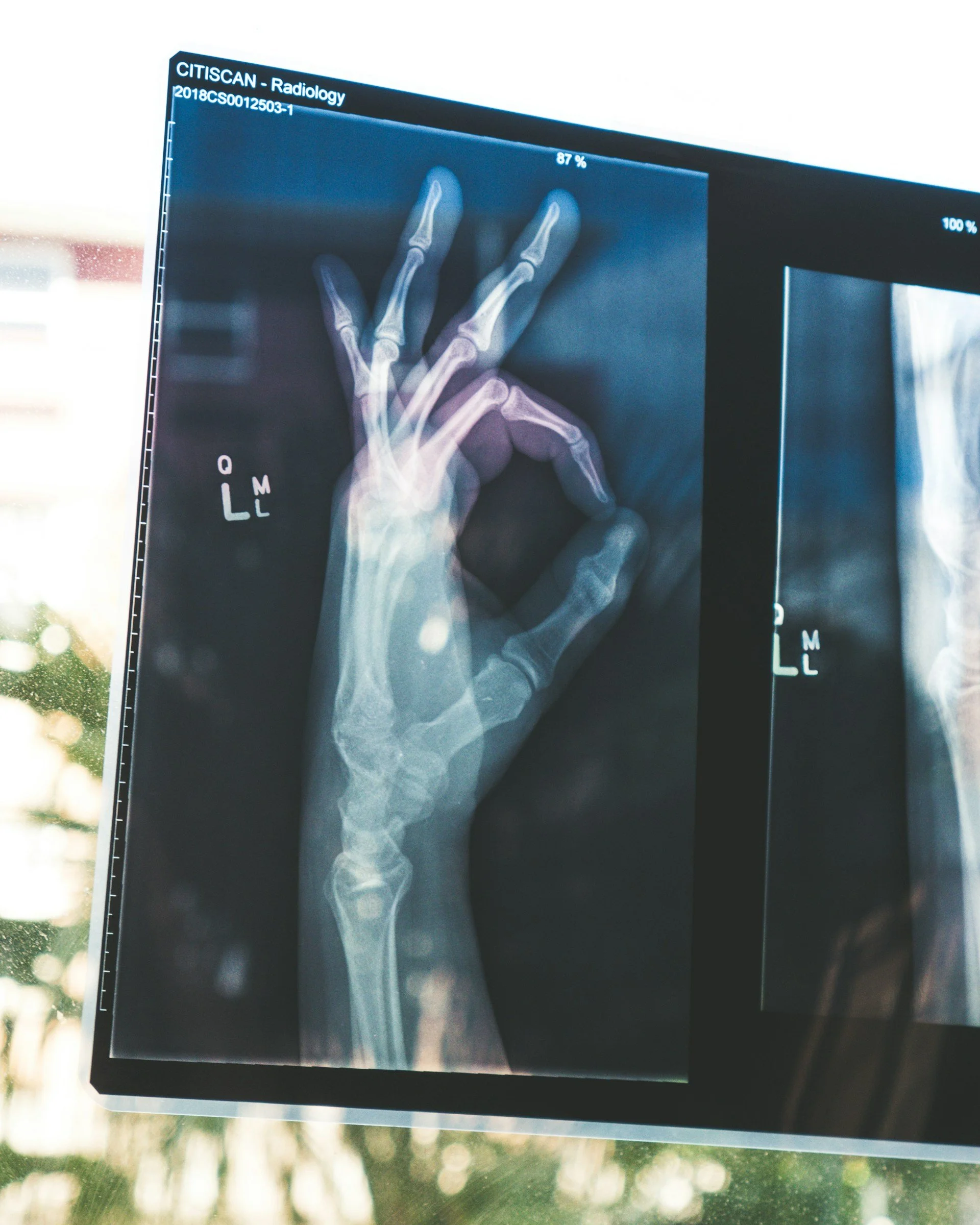
While good optics and imaging in all faculties can be valuable, an image is not always as absolute as the common viewpoint may suggest. Medical imaging such as x-ray, ultrasound scans, MRI, CT and PET scans are adjuncts to diagnosis, monitoring and treating diseases and guiding treatment plans. The clinician should always be more accurate in evaluation than the image.
A large New Zealand insurer engaged a very good medical researcher to see if MRI could be used as a ‘ Gold Standard.’ That answer was, (sic) “ useful in just over 50% of cases referred, but no not a Gold Standard. “ Insurers are not bound by research anymore than medical clinicians and patients are bound by an image. However, leaving in place common usage, even if incorrect, can sometimes be helpful to the bottom line.
For those who still imagine an MRI to be enough, the following two quotes may appeal and awake a Judge’s sense of fairness.
In April 2021, doctors writing in the Australian and New Zealand Journal of Surgery (2) wrote:
“Magnetic resonance imaging (MRI) is commonly used for diagnosis and as a research tool, but its accuracy is questionable.” They further wrote that the goal of their study was to compare the accuracy of knee MRI with clinical (in surgery) assessment for diagnosing meniscal tears and to determine the accuracy of MRI for grading chondral lesions. What did they find? “MRI has relatively poor correlation with arthroscopic findings for grading the chondral damage and was less accurate than clinical assessment for the diagnosis of lateral meniscal tears.” After the MRI only 1 out of 6 received appropriate treatment.
When discussing MRI accuracy or reliability, people will often point to the age of the study. Why? Because there is a belief that technology is moving so rapidly that anything more than a few years, months, or even weeks old is already outdated. That is a misnomer. These results are reflected in recent and current research.
A July 2021 paper published in the BMJ open quality (22) suggested a problem with overreliance of MRIs in the United Kingdom. As you can see from the associated research presented here, a very similar problem to that seen here in the United States. In this study the researchers saw that the musculoskeletal injury or pain was the cause for the largest proportion of general practitioner recommended magnetic resonance imaging (MRI). This comes still with “limited supporting evidence and potential harms from early imaging overuse.”
In other words, too many MRIs, too many unneeded surgeries, too many instances where the patient’s situation was made worse.
Of course the case is not over until there is a final verdict and that is: each case on its merits.
Despite a heavy lean by some insurers it is not fait accompli for patients to accept a denial of insurance, treatment, or compensation, solely based on the medical image. Sometimes even a ‘conclusive‘ image is not relevant a short time later. A patient presented with all signs and symptoms suggesting a fractured wrist. The first x-ray was negative, but the problem persisted. A second x-ray about 10 days later was negative, no fracture. Another week passed where the patient deteriorated and (with some opposition) another x-ray was insisted on, at which point a significant fracture was found — one that, had it been missed, would have severely impacted on the patient’s livelihood and health for years to come. Splintage for many weeks followed by rehabilitation annulled all pain and symptoms. Judgement verse evidence.
Some say humankind is made in God’s image, but that does not make us God.
"Man is made in God's image" (*imago Dei) means humans reflect divine qualities like reason, morality, and relational capacity, not a physical likeness, making them unique, valuable, and representatives of God on Earth, with roles for stewardship and communion, though marred by sin but redeemable in many faiths. It signifies inherent worth, demanding respect and dignity for all people, as they bear a special reflection of their Creator.
While likeness or resemblance does not make us God, increased depth or understanding and insight of the portrait are valuable assets paralleling medical imaging which can be worthwhile assets to augment the treatment plan for each of us. Imaging is not God, or even Gold Standard, the nearest to gold remains The Evaluator who interprets all relevant information and decides the next sentence.